Sunday, June 1, 2014

Renal Pharmacology


Golden Rule: Water tends to follow sodium.


source

Mannitol MOA 

osmotic diuretic, increases tubular fluid osmolarity, producing increased urine flow

Hydrochlorothiazide MOA

inhibits NaCl reabsorption in the early distal tubule

Loop diuretics MOA

inhibits cotransport system (Na+, K+, 2Cl-)  in the thick ascending limb of loop of Henle
*abolishes hypertonicity of medulla, preventing concentration of urine

Acetazolamide MOA

carbonic anhydrase inhibitor
*causes self-limited NaHCO3 diuresis and reductions in total body HCO3- stores
 in the proximal convoluted tubule


Saturday, May 31, 2014

Renal Pathology: Glomerular Diseases

Casts in urine: indicates hematuria/pyuira is of renal (vs. bladder) origin
1. RBC casts
renal: glomerulonephritis, ischemia, or malignant hypertension
bladder: bladder cancer, kidney stones --> hematuria, no casts


2. WBC casts

renal: tubulointerstitial inflammation, acute pyelonephritis, transplant rejection
bladder: acute cystitis --> pyuria, no casts

3. Fatty casts ("oval fat bodies"): nephrotic syndrome
4. Granular ("muddy brown") casts: acute tubular necrosis


5. waxy casts: Advanced renal disease/chronic renal failure


6. hyaline casts: Nonspecific, can be a normal finding


What are the three types of proteinuria?


Overflow proteinuria

Low-molecular weight proteins filtered by the glomerulus are almost entirely reabsorbed in the proximal tubule. With increased low-molecular weight protein production, the amount of filtered protein exceeds tubular reabsorptive capacity, leading to proteinuria. 

Tubular proteinuria

Tubulointerstitial diseases (eg, Fanconi syndrome) can lead to decreased reabsorptive capacity of the proximal tubule and up to 2 g of proteinuria per day because of impaired tubular absorption of filtered albumin, as well as loss of tubular proteins (β2 microglobulin).

Glomerular proteinuria

Glomerular proteinuria is a sensitive marker for glomerular disease. It develops as a result of increased filtration of macromolecules (eg, albumin) across the glomerular capillary wall . source


Glomerular Diseases

source


What are the characteristics of nephrotic syndrome?


  • -massive prOteinuria (>3.5 g/day; "frothy")
  • -hyperlipidemia, hypercholesteremia --> fatty casts
  • -hypoalbuminemia --> pitting edema
  • source
  • -hypogammaglobulinemia --> increased risk of infection
  • -loss of antithrombin III --> hypercoagulable state --> thromboembolism
  • -normal GFR at onset
  • -NO glomerular hypercellularity

source


What are the conditions required to diagnose nephrotic syndrome?


  • proteinuria (>3 g/24 hours)
  • PLUS TWO OF THE FOLLOWING
hypoalbuminemia (<3.5 g/L)
edema
hyperlipidemia

Nephrotic Syndrome:First two are non-immune associated nephrotic syndromes.1) minimal change disease:
light microscopy: normal glomeruli
electron microscopy: foot process efacement

source

Key characteristics: loss of albumin, selective proteinuria [selective (resulting in loss of small MW proteins such as albumin) vs non-selective (with loss of larger MW proteins, such as globulins)]
Triggered: immune stimulus
Population: children
Tx: corticosteroids 
What serious disease may cause minimal change disease?
Hodgkin lymphoma: Hodgkin's lymphoma is characterised by the orderly spread of disease from one lymph node group to another and by the development of system disease with advanced disease. 


What is the most common cause of nephrotic syndrome in adults, especially Hispanic and Black patients?
2) Focal segmental glomerulosclerosis

light microscopy: segmental sclerosis and hyalinosis
electron microscopy: effacement of foot process similar to minimal change disease
Population: adults (#1 nephrotic syndrome to affect adults)
Associations: HIV infection, heroin abuse, massive obesity, interferon tx, and chronic kidney disease
4 Types: 


3) Membranous nephropathy

Light microscopy: diffuse capillary and GBM thickening
source

Electron microscopy: spike and dome appearance with sub epithelial deposits
source

immunofluorescence: granular, Systemic lupus erythematosus (SLE) nephrotic presentation
second most common cause of primary nephritic syndrome in adults.
associations: drugs, infection, SLE, solid tumors

4) Amyloidosis
light microscopy: congo red stain shows apple-green
birefringence under polarized light
associations: Primary= multiple myeloma
secondary = autoimmune such TB and Rheumatoid arthritis

5) Membranoproliferative glomerulonephritis (MPGN)
Type 1 = sub endothelial immune complexes deposits
immunofluorescence = granular
electron microscopy = tram-track w/ GBM splitting caused by mesangial ingrowth
association: HBV, HCV
Type 2 = intramembranous immune complexes: dense deposits
can also be presented as nephritic syndrome
association: C3 nephritic factor

6) Diabetic glomerulonephropathy
nonenzymatic glycosylation --> increase permeability, thickening.
nonenzymatic glycosylation of efferent arterioles --> increase GFR --> mesangial expansion
light microscopy - mesangial expansion, GBM thickening, eosinophilic nodular glomerulosclerosis = kimmelstiel-wilson lesion


Nephritic Syndrome:

What are the symptoms of nephritic syndrome?

  • -hematuria (>5 RBCs per high power field; RBC casts and dysmorphic RBCs)
  • -limited proteinuria (<3.5; note that the protein is not GREATLY elevated and is nonselective)
  • -oliguria and azotemia
  • -impairment of renal function (abrupt increase of creatine, decreased GFR) --> retention of sodium and water --> HTN, edema
  • -hypercellular, inflamed glomeruli
  • -immune-complex deposition --> complement activation --> C5a attraction of neutrophils --> damage
1) Acute Poststreptococcal glomerulonephritis
light microscopy: glomeruli enlarged and hyper cellular neutrophils, "lumpy-bumpy" appearance.
electron microscopy: subepithelial immune complex (IC) humps
immunofluorescence: granular appearance due to IgG, IgM and C3 deposition along GBM and mesangium.
Population: most frequently seen in children. Peripheral and periobital edema, dark urine and hypertension.
prognosis: good. resolves spontaneously
2)rapidly pogressive (crescentric) glomerulonephritis (RPGN): 
light microscopy and immunofluorescence: crescent moon shape. crescents consist of fibrin and plasma protein with glomerular parietal cells, monocytes, and macrophages.
Prognosis: rapidly deteriorating renal function. not good.

  • Goodpasture's syndrome = type ll = hypersensitivity; antibodies to GBM and alveolar basement membrane --> linear IF
  • c-ANCA = Granulomatosis with polyangiitis (Wegener's) 
  • p-ANCA = Microscopic polyangiitis
2) Diffuse proliferative glomerulonephritis (DPGN) 
  • causes: SLE or MPGN
  • light microscopy = wire looping of capillaries

  • EM- sub endothelial and sometimes intrmaembranous IgG-based ICs often with C3 deposition
  • IF - granular
  • most common cause of death = SLE
3) Berger's Disease (IgA Nephropathy)
  • Association: enoch-schonlein purpura
    • Henoch-Schonlein purpura is a disease that involves purple spots on the skin, joint pain, gastrointestinal problems, and glomerulonephritis (a type of kidney disorder).

  • LM - mesangial proliferation
  • EM - mesangial IC deposits
  • IF - IgA based IC deposits in mesangium
  • symptoms: URI or acute gastroenteritis
4) Alphort Syndrome
  • mutation in type 4 collagen --> split basement membrnae
  • mode of inheritance = x linked dominant
  • symptoms: deafness, ocular problems

Wednesday, May 28, 2014

First Aid Renal Physiology Part ll


Nephron Physiology
source


source

Key Points: 

Early Proximal Tubule: reabsorb all glucose, amino acids and most of bicarbonate, sodium, chloride, phosphate and water. Generates and secretes ammonia.
Thin descending loop of Henle: passive reabsorbs water.
Think Ascending Loop of Henle: actively reabsorbs Na+, K+ and Cl-. Responsible for paracellular reabsorption of Mg2+ and Ca2+. Impermeable to H2O.
Early Distal Convoluted Tubule: actively reabsorb Na_ and Cl-.
Collecting Tubule: reabsorb Na+ by secreting K+ and H+ using aldosterone. Reabsorb water by using ADH.

PTH: 
1) at the PCT = inhibits Na+/Phosphate cotransport and promotes phosphate excretion 
2) at the Early distal convoluted tubule = increase Ca2+/Na+ exchange and promotes Ca2_ reabsorption


Relative Concentrations along proximal tubule


Renin -angiotensin-aldosterone system
juxtaglomerular apparatus
source
1) Macula Densa
Osmoreceptor cells in the Distal Convoluted Tubule that monitor osmolarity of filtrate

2) Juxtaglomerular Cells

Enlarged, smooth muscle cells that have secretory granules containing renin. These act on mechanoreceptors.


3) Mesanglial Cells

Have phagocytic and contractile properties and also influences capillary filtration.

OVERVIEW:


Angiotensin ll influences:
1) Baroreceptors: Baroreceptor reflex influences the GFR via long term regulation of arterial blood pressure

1. Higher arterial blood pressure triggers both long term and short term changes to lower blood pressure. 

2. High blood pressure sensed in aortic arch and carotid sinus baroreceptors
3. These release vasoactive chemicals that increase sympathetic activity and promote generalized arteriolar constriction such as ATP, endothelin/ADH/vasopressin, and adenosine
4. The increased sympathetic activity increases cardiac output 
5. Increased sympathetic activity also increases general vasoconstriction - leading to higher total peripheral resistance and afferent arteriolar vasoconstriction
6. Thus, glomerular capillary blood pressure goes down,
7. GFR down
8. Urine volume down
9. More salt and fluid are conserved
10. These go back to increase blood pressure


Thus, low blood pressure promotes long and short term adjustment. Short term is the increased sympathetic activity and vasoconstriction effects to increase cardiac output and total peripheral resistance. Long term is the vasoconstriction of afferent arterioles to glomerulus, lowering GFR, lowering urine volume and retaining fluid and salt to increase blood pressure.


2) AT1 RECEPTOR: vascular smooth muscle vasoconstriction = increase BP


3) ALDOSTERONE: Adrenal gland releases aldosterone.

SOURCE
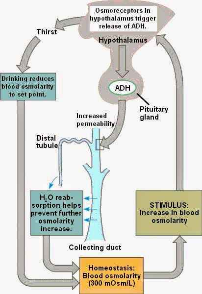
4) ADH: released by the pituitary gland.
SOURCE

Kidney Endocrine Functions: 
1) Erythropoietin released by interstitial cells of the peritubular capillary due to hypoxia.
source
2) 1, 25 - (OH)2 vitamin D
source
  • PTH indirectly stimulates Ca++ absorption in the small intestine by stimulating synthesis of 1,25(OH)2D in the kidney.
  • 25-OH vitamin D is converted to 1,25-(OH)2 vitamin D (the active form) by 1 alpha hydroxylase
3) Renin
Triggers to release renin: decreased BP, low sodium level (macula densa)  and increased sympathetics.
4) Prostaglandins

paracrine secretion vasodilates afferent arterioles. Effect can be inhibited by NSAIDs which then lead to acute renal failure.
source


Hormones acting on kidney

1) artial natiruetic peptide (ANP)
detects increases in BP and responds by prompting the kidney to keep as much H2O as possible in the filtrate. Mechanism: decreasing NaCl reabsorption and therefore decreasing H2O (b/c it follows sodium)


2) Parathyroid Hormone (PTH)


low plasma calcium, high plasma phosphate or low plasma 1,25-(OH)2 vitamin D ===> increases concentration of calcium resorption in DCT, decreases phosphate reabsorption in the PCT, increase 1,25-(OH)2 vitamin D production in the kidney
PTH increases calcium reabsorption in DCT

source


PTH decreases phosphate reabsorption in PCT

source


3) Angiotensin ll (AT ll)
decreased BP ==> efferent arteriole constriction ==> increase GFR and FF (FF = GFR/RPF)
  • Angiotensin II stimulates Na+/H+ exchangers located on the apical membranes (faces the tubular lumen) of cells in the proximal tubule and thick ascending limb of the loop of Henle in addition to Na+ channels in the collecting ducts. This will ultimately lead to increased sodium reabsorption
4) Aldosterone
decrease blood volume + increased plasma osmolarity (K+) ===> aldosterone secretion ==> increased Na+ resorption with K+ and H+ secretion
source

source
5) ADH
increased plasma osmolarity + decreased blood volume ==> ADH secretion ==> ADH bind to principal cells' receptors ==> increase aquaporins ==> increase H2O absorption 
source
Potassium shifts


Shifts K+ out of cell (causing hyperkalemia): DO Insulin LAB
1. Digitalis
source
digitalis inhibits the Na-K ATPase (which pumps 3 Na out of the cell and 2 K in); as such, it can result in hyperkalemia and a variety of cardiac arrhythmias.
2. HyperOsmolarity
hyperosmolarity induces water efflux out of cells, and by solvent drag increases intravascular potassium concentrations. Drugs such as mannitol can therefore cause translocational hyperkalemia.


3. Insulin deficiency
Ingested K+ is absorbed rapidly and enters the portal circulation, where it stimulates insulin secretion. Insulin increases Na+,K+-ATPase activity and facilitates potassium entry into cells, thereby averting hyperkalemia. 
4. Lysis of cells
When there is a rapid amount of cellular destruction, the components of the cells (including potassium), will move outside of the cell, and into the blood stream. 
5. Acidosis 

source
6. B-adrenergic antagonist
Normally, agonist binding to the beta2-adrenergic receptor stimulates the formation of cyclic AMP, which acts through protein kinase A to phosphorylate and activate the Na-K-ATPase pump, leading to the influx of potassium into cells. Competitive inhibition of the beta2 receptor by beta blockers decreases Na-K-ATPase function and reduces potassium uptake by cells.
source


Shifts K+ Into Cell (causing Hypokalemia) Insulin shifts K+ into cells
1) Hypo-osmolarity
2) Insulin 
3) Alkalosis
source


4) B-adrenergic agonist

Electrolyte disturbances
LOW SERUM CONCENTRATION
1) Na+ : nausea, malaise, stupor, coma
hyponatremia

2) K+ : U waves on ECG, flattened T waves, arrhythmias, muscle weakness
hypokalemia
source

3) Ca2+ : Tetany, seizures
hypocalcemia


4) Mg2+ : Tetany, arrhythmias
Hypomagnesiumia 
 hypomagnesemia can cause hypocalcemia since magnesium is necessary for the production of PTH.


PTH
Calcium
Phosphate
Si/sx
Primary Hyperparathyroidism
stones, bones, abdominal groans and psychic moans"
Chronic Renal Failure (→Vit D def.)
↑ - can’t be excreted

Hypercalcemia (malignancy)

Hypomagnesemia
Hypocalcemia that responds to Mg 2+infusion
5) PO43- : bone loss, osteomalacia (bone softening)
Hypophosphatemia

HIGH SERUM CONCENTRATION: 
1) Na+ : irritability, stupor, coma
hypernatremia
2) K+ : Wide QRS and peaked T waves on ECG, arrhythmias, muscle weakness
hyperkalemia
3) Ca2+ Stones, Bones, Grones, and psychiatric overtones
renal STONES
BONE pain
abdominal pain
anxiety, altered mental status
but not necessarily calciuria

source


4) Mg2+
hypermagnesemia
decreased DTRs, lethargy, bradycardia, hypotension, cardiac arrest, hypocalcemia (Mg2+ inhibits calcium from being released from the sarcoplasmic reticulum)
5) PO43-
Renal stones, metastatic calcifications, hypocalcemia

Overview: 
Magnesium (Mg2+) has a direct relationship with potassium (K+) and calcium (Ca2+).
K+ has a direct relationship with Mg2+ and Ca+.
Ca2+ has a direct relationship with K+ and Mg2+.
Mg2+ is needed for K+ absorption.

Calcium has an inverse relationship with phosphate.

Acid-base physiology


What is the Henderson-Hasselbalch equation?pH = 6.1 + log ([HCO3-]/[.03 x PCO2])

What is Winter's formula, and for what is it used? 

APPLICATION calculates predicted respiratory compensation for a simple metabolic acidosis
NOTE if the measured PCO2 differs significantly from that calculated using Winter's formula, then a mixed acid-base disorder is present

Acidemia:
Respiratory acidosis: pH < 7.4, PCo2 > 40 mmHg
causes: airway obstruction, acute lung disease, Chronic lung disease, Opioids, sedatives, weakening respiratory muscles

Metabolic acidosis with compensation (hyperventilation) pH < 7.4, PCo2 < 40 mmHg
1) increased anion gap
MUDPILES
Methanol (formic acid)
Uremiais the illness accompanying kidney failure (also called renal failure), in particular the nitrogenous waste products associated with the failure of this organ.
Diabetic ketoacidosis = develops when your body is unable to produce enough insulin. Insulin normally plays a key role in helping sugar (glucose) — a major source of energy for your muscles and other tissues — enter your cells. Without enough insulin, your body begins to break down fat as an alternate fuel. This process produces a buildup of toxic acids in the bloodstream called ketones, eventually leading to diabetic ketoacidosis if untreated.
Propylene glycol
Iron tablets
Lactic acidosis - The condition typically occurs when cells receive too little oxygen (hypoxia), for example, during vigorous exercise. In this situation, impaired cellular respiration leads to lower pH levels. Simultaneously, cells are forced to metabolize glucose anaerobically, which leads to lactate formation. Therefore, elevated lactate is indicative of tissue hypoxia, hypo perfusion and possible damage.
Ethylene glycol (oxalic acid)
Salicylates (late)

2) Normal anion gap (8-12 mEq/L)
HARD-ASS
Hyperalimentation =  refers to a state where quantities of food consumed are greater than appropriate. It includes overeating, as well as other routes of administration such as in parenteral nutrition.
Addison's disease (adrenal insufficiency) = a rare, chronic endocrine disorder in which the adrenal glands do not produce sufficient steroid hormones (glucocorticoids and often mineralocorticoids).
Renal Tubular Acidosis = a disease that occurs when the kidneys fail to excrete acids into the urine, which causes a person's blood to remain too acidic. 
Diarrhea
Acetazolamide (carbonic anhydrase inhibitor)
 blunts the Na+/H+ exchanger effect. Additionally, HCO3– is retained in the lumen, with marked elevation in urinary pH. The loss of HCO3–causes a hyperchloremic metabolic acidosis 
source


Spironolactone = a competitive antagonist of the aldosterone (or mineralocorticoid) receptor 
Anything that causes your serum to have high K+ will causes all cells to shift K+ into the cell in exchange for H+ out of the cell through the K+/H+ ATPase.

Saline infusion (pH 5.5) - bicarbonate dilution

Alkalemia 
Respiratory alkalosis: pH > 7.4, PCO2 < 40 mmHg
hyperventilation and salicylates (early)
Metabolic alkalosis with compensation (hypoventilation) pH > 7.4, PCO2 > 40  mmHg
Loop diuretics
Loop diuretics block the Na/K/Cl co-transporters in the distal nephron. This causes a decrease in the reabsorption of chloride ions and therefore, increases the luminal electronegativity of the distal nephron. The increase in luminal electronegativity allows excessive 'wasting' of both hydrogen and potassium ions, as these cations will be attracted to the relatively negative charge in the lumen. Over time, the  depletion of cations (particularly hydrogen ions) causes the plasma pH to increase (i.e. alkalosis). source
source
source


Vomiting
Antacid use
source

Hyperaldosteronism : Renal loss of hydrogen ions occurs when large amount of aldosterone increases the activity of a sodium-hydrogen transporter in the kidney. This retains sodium ions whilst pumping hydrogen ions into the renal lumen. The loss of hydrogen ions creates a metabolic alkalosis.